|
X-Ray, CT or Ultrasound: Which Scan Best Detects a Rib Fracture?
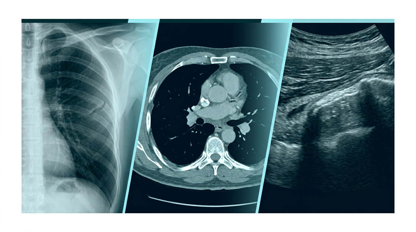
A cracked rib is one of those injuries that sounds minor until the pain of every breath proves otherwise, and the imaging choice made in the emergency department can significantly affect whether the fracture actually gets identified at all.
Not all imaging methods perform equally here, and the gap between them is larger than most patients realise until they're the ones waiting on results.
Why Standard X-Rays Miss So Many Rib Fractures
Traditional chest X-ray has surprisingly poor sensitivity for rib fractures specifically, with some studies citing figures as low as 15% detection for fractures that are later confirmed on CT scanning of the same patient.
The reason is largely anatomical: ribs curve in three dimensions, and a two-dimensional X-ray image can easily miss a fracture line that isn't oriented favourably relative to the angle the image was captured from.
CT Scanning as the More Reliable Standard
A large emergency department study found that of patients who had both a chest CT and a chest X-ray, only 66% of the fractures visible on CT were also seen on the corresponding X-ray, meaning roughly a third went undetected on the more commonly ordered scan.
Trauma centres that see high volumes of chest injuries have increasingly adopted a lower threshold for ordering CT, particularly for patients over sixty or those with more than one suspected fracture site.
Anyone with persistent chest wall pain after trauma, even when an initial scan comes back clear, may benefit from a follow-up CT scan if symptoms don't improve as expected within a week or so, since a missed fracture can complicate pain management and recovery planning.
Dr Marco Scarci give an excellent explanation of what an X-ray of broken ribs shows here: https://marcoscarci.co.uk/x-ray-of-broken-ribs/
Where Ultrasound Fits Into the Picture
Point-of-care ultrasound has emerged as a useful bedside tool, particularly for anterior and lateral rib fractures where the bone sits close enough to the skin surface for sound waves to detect a break in the cortical outline clearly.
Separate research comparing chest radiographs against CT for rib fracture detection found radiologists using CT correctly identified roughly three times as many fractures as those reading X-rays alone, a gap large enough that many trauma centres now default to CT whenever multiple rib fractures are suspected clinically, a finding that reinforces why ultrasound is generally treated as a complementary tool rather than a full CT replacement.
It performs less reliably for posterior rib fractures, where surrounding muscle and the angle of the ribs against the chest wall make visualisation considerably harder, which limits its use as a standalone diagnostic tool for the full rib cage.
Training and equipment availability also shape how consistently ultrasound gets used this way, since the technique depends heavily on operator skill in a way that standard X-ray interpretation generally does not.
Why the Choice Sometimes Comes Down to Practicality, Not Just Accuracy
CT scanning delivers a meaningfully higher radiation dose than a plain X-ray, a real consideration for younger patients or anyone requiring repeated imaging, which is part of why X-ray often remains the first step despite its known limitations.
Emergency departments also weigh cost and availability. Not every facility has a CT scanner accessible around the clock, and for straightforward cases with clear clinical findings, the added precision of CT may not always change the immediate management plan.
Clinicians typically weigh these trade-offs case by case rather than following a rigid protocol, factoring in the mechanism of injury, the patient's age, and whether symptoms suggest a complication like a punctured lung that would justify the additional radiation exposure of CT imaging.
When Missing a Fracture Actually Matters Clinically
For an isolated, uncomplicated rib fracture, treatment is largely supportive, pain control and breathing exercises, regardless of whether the fracture was formally confirmed on imaging, which somewhat reduces the practical stakes of a missed diagnosis in simple cases.
The stakes rise considerably for multiple rib fractures, elderly patients, or anyone with reduced lung function at baseline, where missing the full extent of injury can mean underestimating complication risk, particularly pneumonia, which becomes significantly more common as the number of fractured ribs increases.
Elderly patients with three or more rib fractures face a notably elevated risk of pneumonia and reduced lung capacity during recovery, which is why physicians tend to escalate to CT imaging more readily in this group even when initial X-rays appear reassuring.
For most otherwise healthy adults with an isolated, uncomplicated fracture, the imaging choice matters less for long-term outcome than consistent pain control does, since inadequate pain management itself increases pneumonia risk by discouraging deep breathing and effective coughing during recovery.
The break in the bone itself is thin enough in many early or subtle fractures that even an experienced radiologist reviewing a standard two-view X-ray series can reasonably miss it, which is part of why clinical suspicion should carry real weight alongside whatever the initial imaging shows.
A patient whose pain persists well beyond the typical healing window for a minor fracture, generally four to six weeks, is often worth reassessing regardless of what the original imaging showed at the time of injury.
|